THE CHALLENGE
Lameness is a term typically used to describe a change in a horse gait in response to pain in one of the distal limbs. Sometimes, lameness is obvious manifesting as a noticeable limp; other cases have more subtle signs and can be harder to detect. Successfully resolving lameness cases due to distal limb problems depends mainly on the veterinarian’s skill in diagnosis using both physical exam and more technical diagnostic tools. Currently, most veterinary students train using live animals, which can be dangerous due to the unpredictability of animals in pain. As a result, simulation-based training is essential in veterinary medicine education and practice.
OUR SOLUTION
Fawzy Elnady and his team at Virginia Tech have developed a horse distal limb ultrasound simulator that will enable students to learn and practice ultrasound technique without the risks associated with working with animals. The simulator uses ethically-sourced equine distal limbs preserved using the Elnady technique with RFID chips embedded at key locations. As students move an ultrasound wand over the limb, videos of ultrasound recordings will play when the wand is properly positioned. Instructors have the ability to choose between normal imaging or ultrasounds indicating pathologies. The simulator limbs are dry, flexible, odorless, and have high fidelity to real limbs.
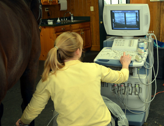
Figure: Detailed images produced by diagnostic ultrasound can help evaluate injuries and inflammation in tendons, ligaments and other soft tissues. (Photo from Virginia Equine Imaging via Practical Horseman)